Nervous Tissue
- Nervous Tissue
Although the nervous system is very complex, there are only two main types of cells in nervous tissue. Nervous tissue is the term for groups of organized cells in the nervous system, which is the organ system that controls the body’s movements, sends and carries signals to and from the different parts of the body, and has a role in controlling bodily functions such as digestion. Nervous tissue is grouped into two main categories: neurons and neuroglia. Neurons, or nerves, transmit electrical impulses, while neuroglia do not; neuroglia have many other functions including supporting and protecting neurons.
Nervous tissues are made of cells specialized to receive and transmit electrical impulses from specific areas of the body and to send them to specific locations in the body organized into structures called nerves. A nerve consists of a neuron and glial cells. The main cell of the nervous system is the neuron.
There is a large structure with a central nucleus: the cell body (or soma) of the neuron. Projections from the cell body are either dendrites, specialized in receiving input, or a single axon, specialized in transmitting impulses. Glial cells support the neurons. Astrocytes regulate the chemical environment of the nerve cell, while oligodendrocytes insulate the axon so the electrical nerve impulse is transferred more efficiently. Other glial cells support the nutritional and waste requirements of the neuron. Some of the glial cells are phagocytic, removing debris or damaged cells from the tissue.
- Neurons
Your brain is made up of millions of cells called neurons. Neurons make connections with each other to create pathways that control all aspects of life, such as bodily functions, emotions, and movement. Each neuron is composed of three parts: the cell body, dendrites, and an axon.
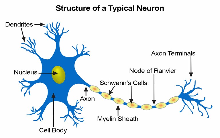
Neurons, or nerve cells, carry out the functions of the nervous system by conducting nerve impulses. They are highly specialized and amitotic. This means that if a neuron is destroyed, it cannot be replaced because neurons do not go through mitosis. The image below illustrates the structure of a typical neuron.
Each neuron has three basic parts: cell body (soma), one or more dendrites, and a single axon.
- Cell Body
The cell body, also called the soma, is the spherical part of the neuron that contains the nucleus. The cell body connects to the dendrites, which bring information to the neuron, and the axon, which sends information to other neurons. When information is received from another neuron, the dendrites pass the signal to the cell body. The cell body then may send the information to the axon, depending on the strength of the signal.
In many ways, the cell body is similar to other types of cells. It has a nucleus with at least one nucleolus and contains many of the typical cytoplasmic organelles. It lacks centrioles, however. Because centrioles function in cell division, the fact that neurons lack these organelles is consistent with the amitotic nature of the cell.
- Dendrites
Dendrites and axons are cytoplasmic extensions, or processes, that project from the cell body. They are sometimes referred to as fibers. Dendrites are usually, but not always, short and branching, which increases their surface area to receive signals from other neurons. The number of dendrites on a neuron varies. They are called afferent processes because they transmit impulses to the neuron cell body. There is only one axon that projects from each cell body. It is usually elongated and because it carries impulses away from the cell body, it is called an efferent process.


